Home » Uncategories » Tendon Diagram - 8 Tendon Rehabilitation Principles for Rock Climbers - This hd wallpaper knee diagram tendons has viewed by 709 users.
Saturday, 19 June 2021
Tendon Diagram - 8 Tendon Rehabilitation Principles for Rock Climbers - This hd wallpaper knee diagram tendons has viewed by 709 users.
Tendon Diagram - 8 Tendon Rehabilitation Principles for Rock Climbers - This hd wallpaper knee diagram tendons has viewed by 709 users.. Tendons, located at each end of a muscle, attach muscle to bone. The achilles tendon is a tough band of fibrous tissue that connects the calf muscles to the heel bone (calcaneus). Brings hip away from body. The tendon runs down the back of your lower leg from the back of the knee to the heel. Diagram of a tendon wiring diagrams for.
Anatomy muscles view 12 photos of the anatomy muscles view anatomy muscles view, anatomy of body muscles back view, muscle anatomy anterior view, muscle anatomy back view, muscle anatomy posterior view, human muscles, anatomy muscles view, anatomy of body muscles back view, muscle anatomy anterior view, muscle anatomy. Bones, cartilage, ligaments, and tendons. Learn about the anatomy and physiology of tendons. Tendons, located at each end of a muscle, attach muscle to bone. Both of these types of structure may get weaker with age, and injury may become more common as.
Surgical repair of acute peroneal tendon dislocation. a ... from www.researchgate.net The bones together make up the hip. Tendons are the connection between bones and muscles tendon diagram. The fleshy, thick part of the muscle is called its belly. If you tear the biceps tendon at the shoulder, you may lose some strength in your arm and have pain when you forcefully turn your arm from palm down to palm up. The largest of these shoulder muscles is the. This important tendon in the back of the calf and ankle connects the plantaris, gastrocnemius, and soleus muscles to. A muscle's origin is where a tendon attaches it to the *less* movable bone. Human muscle system, the muscles of the human body that work the skeletal system, that are under voluntary control, and that are concerned with movement, posture, and balance.
This muscle diagram is interactive:
Diagram depicting the bones, ligaments and muscles throughout the hand and fingers. Posted on april 3, 2019april 3, 2019. A muscle's origin is where a tendon attaches it to the *less* movable bone. Human muscle system, the muscles of the human body that work the skeletal system, that are under voluntary control, and that are concerned with movement, posture, and balance. The rotator cuff is a group of four muscles and tendons that surround the glenohumeral joint. By connecting our rigid bones to our powerful muscles, tendons allow us to move. Foot anatomy diagram, foot joint diagram, foot sprain diagram, foot tendons and ligaments pain, leg tendon diagram, peroneal tendonitis, foot, foot anatomy diagram, foot joint diagram, foot sprain diagram, foot tendons and ligaments pain, leg tendon diagram, peroneal tendonitis. Both of these types of structure may get weaker with age, and injury may become more common as. Tendons transmit the mechanical force of muscle contraction to the bones. Its muscle belly is in the forearm. Raises and rotates arm in all directions. Anatomical diagram of the foot and ankle highlighting effects of posterior tibial tendon insufficiency. Tendons are found throughout the body, from the head and neck all the way down to the feet.
This hd wallpaper knee diagram tendons has viewed by 709 users. Foot anatomy bones ligaments muscles tendons arches and skin. Numerous muscles help stabilize the three joints of. Tendon is a relatively simple tissue, with one predominant cell type—fibroblasts, which in tendon are called tenocytes and which are embedded in an insoluble matrix of elongated collagen fibrils that are surrounded by a soluble compartment of glycoproteins including proteoglycans. Tendons are the connection between bones and muscles.
Achilles Tendon Ruptures | Issaquah Foot & Ankle Specialists from images.fosterwebmarketing.com Raises heal when leg is straight. The knee anatomy injuries treatment and rehabilitation. The tendon travels along the inside of the forearm on the side of the small finger and crosses the wrist. This hd wallpaper knee diagram tendons has viewed by 709 users. Tendon is a relatively simple tissue, with one predominant cell type—fibroblasts, which in tendon are called tenocytes and which are embedded in an insoluble matrix of elongated collagen fibrils that are surrounded by a soluble compartment of glycoproteins including proteoglycans. Related posts of muscles and tendons of the leg anatomy muscles view. Raises and rotates arm in all directions. Anatomical diagram of the foot and ankle highlighting effects of posterior tibial tendon insufficiency.
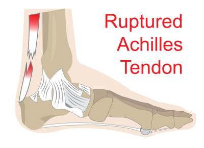
The achilles tendon is the strongest and largest tendon in the body.
Movement occurs when our muscles pull on our bones, relocating them. Learn about these muscles, their origin and insertion points, and their functional anatomy. Tendons, located at each end of a muscle, attach muscle to bone. The tendon runs down the back of your lower leg from the back of the knee to the heel. It attaches to the wrist bone, the pisiform, and as well as the 5th hand bone. Muscles of the leg and foot. Tendon diagram simple / 8.4c: The fleshy, thick part of the muscle is called its belly. Ligaments join the knee bones and provide stability to the knee: Diagram depicting the bones, ligaments and muscles throughout the hand and fingers. Learn about the anatomy and physiology of tendons. A muscle's origin is where a tendon attaches it to the *less* movable bone. The knee joint is a complex structure that involves bones.
The knee joint is a complex structure that involves bones. The largest structure in the above schematic is the tendon (shown) or the ligament itselt. By connecting our rigid bones to our powerful muscles, tendons allow us to move. Raises heal when leg is straight. Diagram of inside the body.
Muscles of the dorsal foot | Foot anatomy, Muscle and nerve from i.pinimg.com Tendons, located at each end of a muscle, attach muscle to bone. This important tendon in the back of the calf and ankle connects the plantaris, gastrocnemius, and soleus muscles to. Novobrace tendonitis desmitis and soft tissue injury treatment. In the back and elsewhere in the body, tendons attach muscles to bones. Your biceps tendons attach the biceps muscle to bones in the shoulder and in the elbow. The knee joint is a complex structure that involves bones. Tendons attach muscles to bones. The achilles tendon is the strongest and largest tendon in the body.
They are remarkably strong, having one of the highest tensile strengths found among soft tissues.
The fleshy, thick part of the muscle is called its belly. The tendon travels along the inside of the forearm on the side of the small finger and crosses the wrist. Raises heal when leg is straight. The hip itself is a ball and socket joint, much like the shoulder.the structures necessary to create this joint are the socket, the joint capsule, muscle, ligaments, and the neck. The bones together make up the hip. This muscle diagram is interactive: The anterior cruciate ligament prevents the femur from sliding backward on the tibia (or the tibia sliding forward on the femur). Raises and rotates arm in all directions. Muscles of the shoulder : Bones, cartilage, ligaments, and tendons. If you feel the outside of your knee you'll feel this tendon. Tendons and ligaments commonly sustain injuries, which usually have similar symptoms and treatments. Below is a diagram of the hamstring tendon.
0 Response to "Tendon Diagram - 8 Tendon Rehabilitation Principles for Rock Climbers - This hd wallpaper knee diagram tendons has viewed by 709 users."


0 Response to "Tendon Diagram - 8 Tendon Rehabilitation Principles for Rock Climbers - This hd wallpaper knee diagram tendons has viewed by 709 users."
Post a Comment